Copaxone: yet another mechanism

Copaxone: yet another mechanism

Previously on this blog I've written about Copaxone and its "mechanism of the week". For example, here and here. I haven't done it for a while, but can't resist bringing this one to your attention.

Now it's anti-bacterial. What next? It inhibits EBV :-)

But I guess it must be working by blocking the microbiome:-).


Christiansen SH, Murphy RA, Juul-Madsen K, Fredborg M, Hvam ML, Axelgaard E, Skovdal SM, Meyer RL, Sørensen UBS, Möller A, Nyengaard JR, Nørskov-Lauritsen N, Wang M, Gadjeva M, Howard KA, Davies JC, Petersen E, Vorup-Jensen T. The Immunomodulatory Drug Glatiramer Acetate is Also an Effective Antimicrobial Agent that Kills Gram-negative Bacteria.
Sci Rep. 2017; 7(1):15653. doi: 10.1038/s41598-017-15969-3.

Classic drug development strategies have failed to meet the urgent clinical needs in treating infections with Gram-negative bacteria. Repurposing drugs can lead to timely availability of new antibiotics, accelerated by existing safety profiles. Glatiramer acetate (GA) is a widely used and safe formulation for treatment of multiple sclerosis. It contains a large diversity of essentially isomeric polypeptides with the cationic and amphiphilic character of many antimicrobial peptides (AMP). Here, we report that GA is antibacterial, targeting Gram-negative organisms with higher activity towards Pseudomonas aeruginosa than the naturally-occurring AMP LL-37 in human plasma. As judged from flow cytometric assays, bacterial killing by GA occurred within minutes. Laboratory strains of Escherichia coli and P. aeruginosa were killed by a process of condensing intracellular contents. Efficient killing by GA was also demonstrated in Acinetobacter baumannii clinical isolates and approximately 50% of clinical isolates of P. aeruginosa from chronic airway infection in CF patients. By contrast, the Gram-positive Staphylococcus aureus cells appeared to be protected from GA by an increased formation of nm-scale particulates. Our data identify GA as an attractive drug repurposing candidate to treat infections with Gram-negative bacteria.


And whilst on the subject:


Grossman I, Kolitz S, Komlosh A, Zeskind B, Weinstein V, Laifenfeld D, Gilbert A, Bar-Ilan O, Fowler KD, Hasson T, Konya A, Wells-Knecht K, Loupe P, Melamed-Gal S, Molotsky T, Krispin R, Papir G, Sahly Y, Hayden MR. Compositional differences between Copaxone and Glatopa are reflected in altered immunomodulation ex vivo in a mouse model. Ann N Y Acad Sci. 2017 1407(1):75-89.


Glatiramer acetate is big business as it is the number one selling drug. But there's one problem: (no, not its level of efficacy) Copaxone is now out of patent protection and generics are entering the market place. 

So what do you do to protect your product?


You aim to show that the competitor compound is different?


Copaxone is a random mix of 4 amino acids and so different batches of the drug are different. 


Glatopa (a generic Copaxone that induces some genes different to Copaxone) is one variant, but is it therapeutically different?


Can coffee really be good for you?

Can coffee really be good for you?

As you see we are trying to post less often on the blog so that you have more time to read and discuss the posts. The following blog post is a bit of a cheat as I have reviewed three papers. However, they are all related to coffee and are relevant to pwMS and their families.

Are you addicted to coffee? If you are, don't worry, the latest research suggests it may be good for you. However, if you have urinary symptoms, anxiety, tremor or sleep problems there may be a downside. 


The good news is that coffee consumption seems safe. The study published last week in the BMJ indicates a reduction in the risk of various health outcomes at three to four cups a day. These results don't necessarily mean that coffee consumption is causal; i.e. in itself responsible for the reduction. Coffee drinkers may be different to non-coffee drinkers and this rather than the coffee per se may be the reason for the reduction in risk. The evidence suggests that coffee could be tested as an intervention without significant risk of causing harm in the disease studied. This analysis did not cover MS. Maybe MS is too uncommon to have been noticed by this research group? 


However, a Swedish study has looked at coffee consumption and MS risk. If you are a coffee drinker it appears you reduce your risks of getting MS by ~30% compared to non-coffee drinkers. Again is this simply an association or causation? Is there something that coffee-drinkers do those non-coffee drinkers don't that protects them from getting MS (association)? Or, do the genetic factors that increase your risk of getting MS simply increase your affinity for coffee, and your chances of becoming addicted to coffee (association)? Or is there something in coffee that alters the immune system and reduces your risk of getting MS (causation)? If the observation is causal what is it in coffee that reduces your chances of getting MS? Could it be caffeine? If it is caffeine then other caffeinated drinks should also reduce your risks of getting MS; e.g. tea, energy drinks, cola, etc. Apart from being a stimulant, we know that caffeine has many biological effects including immunological and neuroprotective effects.

Coffee not only reduces your chances of getting MS but is protective for Parkinson's and Alzheimer's disease as well. The latter observations alone make it worthwhile taking up the habit. Which is the reason I give for my habit of drinking 6-8 espresso shots per day. Did you know that on a global level caffeine is the most prevalent human addiction? Could coffee be another lifestyle factor to take into account when optimising your brain health? 

There are downsides to caffeine if you have MS. Too much can make anxiety worse and could exacerbate tremor. If you take too much caffeine, particularly late in the day, it can cause insomnia. This is why I have my last coffee no later than ~3pm. Caffeine is also a mild diuretic and causes increased urine production (see paper 3 below). This may exacerbate bladder problems. I have many patients who report that just one coffee affects their bladder function (urinary urgency and frequency).

How much coffee do you have? Does it help you? 


Coffee and general health:

Poole et al. Coffee consumption and health: umbrella review of meta-analyses of multiple health outcomes. BMJ 2017;359:j5024.

Objectives: To evaluate the existing evidence for associations between coffee consumption and multiple health outcomes.

Design: Umbrella review of the evidence across meta-analyses of observational and interventional studies of coffee consumption and any health outcome.

Data sources: PubMed, Embase, CINAHL, Cochrane Database of Systematic Reviews, and screening of references.

Eligibility criteria for selecting studies: Meta-analyses of both observational and interventional studies that examined the associations between coffee consumption and any health outcome in any adult population in all countries and all settings. Studies of genetic polymorphisms for coffee metabolism were excluded.

Results: The umbrella review identified 201 meta-analyses of observational research with 67 unique health outcomes and 17 meta-analyses of interventional research with nine unique outcomes. Coffee consumption was more often associated with benefit than harm for a range of health outcomes across exposures including high versus low, any versus none, and one extra cup a day. There was evidence of a non-linear association between consumption and some outcomes, with summary estimates indicating largest relative risk reduction at intakes of three to four cups a day versus none, including all cause mortality (relative risk 0.83, 95% confidence interval 0.83 to 0.88), cardiovascular mortality (0.81, 0.72 to 0.90), and cardiovascular disease (0.85, 0.80 to 0.90). High versus low consumption was associated with an 18% lower risk of incident cancer (0.82, 0.74 to 0.89). Consumption was also associated with a lower risk of several specific cancers and neurological, metabolic, and liver conditions. Harmful associations were largely nullified by adequate adjustment for smoking, except in pregnancy, where high versus low/no consumption was associated with low birth weight (odds ratio 1.31, 95% confidence interval 1.03 to 1.67), preterm birth in the first (1.22, 1.00 to 1.49) and second (1.12, 1.02 to 1.22) trimester, and pregnancy loss (1.46, 1.06 to 1.99). There was also an association between coffee drinking and risk of fracture in women but not in men.

Conclusion: Coffee consumption seems generally safe within usual levels of intake, with summary estimates indicating largest risk reduction for various health outcomes at three to four cups a day, and more likely to benefit health than harm. Robust randomised controlled trials are needed to understand whether the observed associations are causal. Importantly, outside of pregnancy, existing evidence suggests that coffee could be tested as an intervention without significant risk of causing harm. Women at increased risk of fracture should possibly be excluded.

Coffee and MS Risk:

Hedström et al. High consumption of coffee is associated with decreased multiple sclerosis risk; results from two independent studies. J Neurol Neurosurg Psychiatry doi:10.1136/jnnp-2015-312176

Objective: Previous studies on consumption of caffeine and risk of multiple sclerosis (MS) have yielded inconclusive results. We aimed to investigate whether consumption of coffee is associated with risk of MS.

Methods: Using two population-representative case–control studies (a Swedish study comprising 1620 cases and 2788 controls, and a US study comprising 1159 cases and 1172 controls), participants with different habits of coffee consumption based on retrospective data collection were compared regarding risk of MS, by calculating ORs with 95% CIs. Logistic regression models were adjusted for a broad range of potential confounding factors.

Results: Compared with those who reported no coffee consumption, the risk of MS was substantially reduced among those who reported a high consumption of coffee exceeding 900 mL daily (OR 0.70 (95% CI 0.49 to 0.99) in the Swedish study, and OR 0.69 (95% CI 0.50 to 0.96) in the US study). Lower odds of MS with increasing consumption of coffee were observed, regardless of whether coffee consumption at disease onset or 5 or 10 years prior to disease onset was considered.
Conclusions: In accordance with studies in animal models of MS, high consumption of coffee may decrease the risk of developing MS. Caffeine, one component of coffee, has neuroprotective properties, and has been shown to suppress the production of proinflammatory cytokines, which may be mechanisms underlying the observed association. However, further investigations are needed to determine whether exposure to caffeine underlies the observed association and, if so, to evaluate its mechanisms of action.

Caffeine and urinary symptoms:

Bradley et al. Evidence of the Impact of Diet, Fluid Intake, Caffeine, Alcohol and Tobacco on Lower Urinary Tract Symptoms: A Systematic Review. J Urol. 2017 Nov;198(5):1010-1020.

PURPOSE: Diet, fluid intake and caffeine, alcohol and tobacco use may have effects on lower urinary tract symptoms. Constructive changes in these modifiable nonurological factors are suggested to improve lower urinary tract symptoms. To better understand the relationship between nonurological factors and lower urinary tract symptoms, we performed a systematic literature review to examine, grade and summarize reported associations between lower urinary tract symptoms and diet, fluid intake and caffeine, tobacco and alcohol use.

MATERIALS AND METHODS: We performed PubMed® searches for eligible articles providing evidence on associations between 1 or more nonurological factors and lower urinary tract symptoms. A modified Oxford scale was used to grade the evidence.

RESULTS: We reviewed 111 articles addressing diet (28 studies), fluid intake (21) and caffeine (21), alcohol (26) and tobacco use (44). The evidence grade was generally low (6% level 1, 24% level 2, 11% level 3 and 59% level 4). Fluid intake and caffeine use were associated with urinary frequency and urgency in men and women. Modest alcohol use was associated with decreased likelihood of benign prostatic hyperplasia diagnosis and reduced lower urinary tract symptoms in men. Associations between lower urinary tract symptoms and ingestion of certain foods and tobacco were inconsistent.

CONCLUSIONS: Evidence of associations between lower urinary tract symptoms and diet, fluid intake and caffeine, alcohol and tobacco use is sparse and mostly observational. However, there is evidence of associations between an increased fluid and caffeine intake and urinary frequency/urgency, and between modest alcohol intake and decreased benign prostatic hyperplasia diagnosis and lower urinary tract symptoms. Given the importance of these nonurological factors in daily life, and their perceived impact on lower urinary tract symptoms, higher quality evidence is needed.
Alemtuzumab hair today gone tommorrow

Alemtuzumab hair today gone tommorrow

NeuroDoc Gnanapavan I didn't sign-up for this!


 I want my hair back ;-(


Front Neurol. 2017 Oct 30;8:569. doi: 10.3389/fneur.2017.00569. eCollection 2017.

Alopecia Universalis following Alemtuzumab Treatment in Multiple Sclerosis: A Barely Recognized Manifestation of Secondary Autoimmunity-Report of a Case and Review of the Literature.

Zimmermann J, Buhl T, Müller M.

Secondary autoimmunity is the most frequent adverse event occurring in almost every other alemtuzumab-treated multiple sclerosis patient. We report a case of a patient with relapsing-remitting multiple sclerosis who reported smooth, circular areas of complete hair loss on both thighs 6 months after the second treatment cycle with alemtuzumab. The patient was diagnosed as having alopecia areata (AA). Within 3 months, AA progressed to complete loss of all body hair (alopecia universalis). Current literature rarely connects alemtuzumab with the onset of alopecia of autoimmune origin. Here, we report a little-noticed autoimmune disease affecting the skin, very likely being associated with alemtuzumab. We emphasize the necessity of careful clinical surveillance of alemtuzumab-treated patients for yet undescribed autoimmune diseases.

Alopecia areata

Alemtuzumab (aka Campath-1H) is arguably one of the biggest early success stories in MS, but as with every cloud that has a silver lining, every silver lining also has a cloud - namely the autoimmune side effects. In the horizon, ocrelizumab and cladrabine loom as omnipresent threats; which makes acceptance of this very unpalatable cloud by the MS community essential. 

We know that reason for the autoimmune disorders following alemtuzumab is a consequence of B-cell hyper-population in the relative absence of a T cell regulation during the period of immune reconstitution. The ability to predict who develops what, however, have been largely unsuccessful to date, and yet with increased exposure to the drug, the types of autoimmune disorders described is expanding.

So what autoimmune disorders have been linked to alemtuzumab?
  • Autoimmune thyroid disease = 30%
  • Immune thrombocytopenic purpura (low platelets) = 1-2%
  • Goodpasture's disease (kidney disease) = <1%
  • Guillain-Barre syndrome (described in the cancer label)
Around 47% exposed to alemtuzumab develop another autoimmune disorder other than their MS. Zimmerman et al. now report a case of alopecia (or hair loss), which may possibly be linked to alemtuzumab. They also point out the alopecia is not unique to alemtuzumab, and has also been reported with teriflunomide and mitoxantrone. In their case, even after cessation of treatment, there was no regrowth of hair at the 6-month follow up.

The EMA (European Medicines Agency) label does report alopecia under side effects of >0.5% in alemtuzumab 12mg treated subjects. Alopecia areata is thought to be T cell (CD8+NKG2D+) mediated damage of the hair follicles, although there may be some B cell involvement here too and has been linked to thyroid autoimmunity.

The authors' advice that those receiving alemtuzumab examine themselves on regular basis for any early signs of hair loss so that prompt steroid treatment can be started.

Training the next generation of UK's MSologists

Tony Blair is famously quoted as saying 'Education, education, education, ...' and putting education at the centre of New Labour's manifesto in 1997. However, is it the type or the quality of education that makes the difference and changes the world?


We are trying to change the world of MS treatment.


I have learned that information in itself may pique attention, but it rarely changes behaviour.

I have been very impressed by the feedback we have had from our initial two MS Academies. Healthcare delegates participate in a 3-day course. They then go to a research or service development project or even an audit in relation to MS. The act of going away and doing something active makes delegates think about their MS practice and consider the service(s) they provide for their patients with MS. In other words, the doing induces a change in behaviour and complements the educational element of the course in changing behaviour.

Doing makes a difference!

If you are interested in joining the MS Academy and attending an MS Masterclass please register your interest via the MS Academy website. Please note these courses are open to non-UK clinicians and having HCPs from other countries helps NHS employees. After all aren't we still European, or even better citizens of the world?


CoI: At present, the Neurology Academy is supported by an unrestricted educational grant from Biogen. Biogen has no input into the design and running of the Academy that is based on the very influential Parkinson's Disease Academy, which has been running for more than a decade. We are also looking for additional sponsors.  
Study shows a B cell depleting treatment is effective

Study shows a B cell depleting treatment is effective

A new study tests a B cell depleting treatment in MS. The results are interesting.


Anti-CD19 depletion kills off most B cell types, except the plasma cell (antibody secreting cell).

So when an anti-CD20, anti B cell treatment (i.e. ocrelizumab) inhibits MS, the T cellers say it is killing the 5-20% of CD20 positive T cells which contain the pathogenic cell population.

This flimsy argument accepts an unlikely event, whilst dismissing
the fact that marked CD4 T cell depletion had limited effect in MS and marked CD8 depletion with DMF (which is a moderate memory B cell depleter) is only moderately effective.

So with this new study it should now be even easier to find the pathogenic T cells, as there are very few CD19, CD3 T cells (>1%).

What do you think? 

Are the T cellers trying too hard?

The only plausible argument of an important T cell action is that B cell depletion is blocking a critical antigen presenting cell function to T cells in humans.

Agius MA, Klodowska-Duda G, Maciejowski M, Potemkowski A, Li J, Patra K, Wesley J, Madani S, Barron G, Katz E, Flor A.
Safety and tolerability of inebilizumab (MEDI-551), an anti-CD19 monoclonal antibody, in patients with relapsing forms of multiple sclerosis: Results from a phase 1 randomised, placebo-controlled, escalating intravenous and subcutaneous dose study.Mult Scler. 2017 Nov 1:1352458517740641. 

BACKGROUND: B cells may be involved in the pathophysiology of multiple sclerosis (MS). Inebilizumab (formerly MEDI-551) binds to and depletes CD19+ B cells.
OBJECTIVES: To assess safety, tolerability, pharmacokinetics, pharmacodynamics and immunogenicity of inebilizumab in adults with relapsing MS.
METHODS: This phase 1 trial randomised 28 patients 3:1 (21, inebilizumab; 7, placebo) to inebilizumab (2 intravenous (IV) doses, days 1 and 15: 30, 100 or 600 mg; or single subcutaneous (SC) dose on day 1: 60 or 300 mg) or matching placebo, with follow-up until at least week 24 or return of CD19+ B-cell count to ⩾80 cells/µL.
RESULTS: Complete B-cell depletion was observed across all doses. Infusion/injection (grade 1/2) reactions occurred in 6/15 patients receiving inebilizumab IV, 2/5 placebo IV and 1/6 inebilizumab SC. Serious adverse events occurred in three patients receiving inebilizumab: pyrexia, mixed-drug intoxication (unrelated to inebilizumab; resulted in death) and urinary tract infection. Mean number of cumulative new gadolinium-enhancing lesions over 24 weeks was 0.1 with inebilizumab versus 1.3 with placebo; mean numbers of new/newly enlarging T2 lesions were 0.4 and 2.4, respectively.
CONCLUSION:Inebilizumab had an acceptable safety profile in relapsing MS patients and showed a trend in reductions in new/newly enlarging and gadolinium-enhancing lesions.

Are we Overdosing Ocrelizumab

The eagles (ocrelizumab for RRMS and PPMS)  may have landed in Europe, but how often should the birds be used to kill cells in MS?

A view-point argues it's too often and that it can be personalised and smart.  I agree but for different reasons.....

Avasarala J. Anti-CD20 Cell Therapies in Multiple Sclerosis-A Fixed Dosing Schedule for Ocrelizumab is Overkill. Drug Target Insights. 2017 Oct 25;11:1177392817737515.
Anti-CD 20 therapies have found significant uses in multiple sclerosis (MS). Based singularly on the accumulated evidence with the use of rituximab (RTX; Rituxan, Genentech, and Biogen) in neuroimmunological diseases, ocrelizumab (OCR; Ocrevus, Genentech) was developed as a treatment option for MS and selectively targets CD20 B cells, a cell surface antigen found on pre-B cells, mature, and memory B cells, but not on lymphoid stem cells and plasma cells. On the basis of indirect evidence, elimination of the antigen-presenting capabilities and antigen nonspecific immune functions of B cells appear to be central to the therapeutic efficacy of anti-CD20 B-cell therapies. An important question is this-Why does the drug need to be dosed at fixed intervals and not based on a measurable endpoint, such as tracking peripheral CD20 cell counts? There is minimal scientific validity in infusing the drug every 6 months particularly if CD20 cell counts are negligible in the peripheral blood. In this analysis, a case is made for following CD19 cell populations as a surrogate for CD20 cells on a monthly basis to guide OCR redosing parameters and does not follow a scheduled dosing parameter.


Ocrelizumab is used every 6 months, but this study argues that this is too often and that it should be personalised based on monitoring CD19 positive B cells so if CD19 positive cells in an individual have not returned you should wait until your next dosing. This is personalised medicine.

Unfortunately there is often no insight provided to the action of ocrelizumab and this is because of the T cell MS world view and all too often the B cell is seen and shown as a single population.
But once you realize this is not true...the pennies can drop (realise the true situation).

We have made the point that cells within the memory B cell populations are the important subtype(s) to target

Memory B Cells are Major Targets for Effective Immunotherapy in Relapsing Multiple Sclerosis.  https://www.ncbi.nlm.nih.gov/ pubmed/28161400

If you monitor this subset to determine retreatment it may become even more personalised and as these cells take a long long time to re-appear (months-years). Retreatment may be even less frequent.

Looking at the graph shown below where doses would be at week 0 and week 24 you can see total B cells are well down for months

So should you dose every 6 months?

I suspect Pharma and Neuros will have their fingers in their ears going La, La, La, La, La in the response to the idea of personalised medicine...I could be wrong

They will give the drug every 6 months and they will follow the label.

This is what the data from the trials indicates them to do and that is the label.

However, if  we have a death (based on events in Lupus and Arthritis where termination of drug development occurred) because of severe infection due to cell depletion, I suspect that people will "pull their fingers out" and take notice of the views above and below. 

There will of course be deaths in people taking drug, because this happens as part of life and not all deaths are drug related. Adverse events could occur early or late  (We are aware of some rumors) this will be due to chance of when the infections strike and there will be infections associated with B cell depletion. But if you take the treatment forever the risks must accumulate due to chance

However, I have hypothesised that ocrelizumab is an induction therapy just like alemtuzumab and cladribine.....Why?

Because, in my current view (this could change) they work the same way and all deplete memory B cells

Based on the unpublished, (why was it not published...Is it ethical to put people through these trials and not properly document the data but in the public domain see the Stephen Hauser ECTRIMS talk or [Click here]), ocrelizumab phase II extension data set show activity in most people 18 months after the last dose.

Does it need a tragedy, before people think about this? 
Maybe the manufacturers should plan and do a trial now...Maybe I am wrong and they can shut me up! 

They can do this by showing us the data.

Personalising Treatment is surely a positive move.

COI: Non-relevant yet.
******************************************************
EDUCATION:


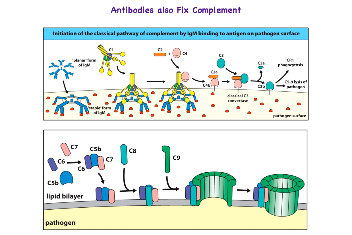
Ocreliziumab is a CD20-killer and it does this by complement fixation to blow holes in the target and uses antibody dependent cellular cytotoxicity to blow holes in the target. 

"Complement fixation" is where the complement binding site on the antibody molecule stimulates the complement cascade starting with C1 and ending with C9.


The complement system consists of a number of small proteins found in the blood, in general synthesized by the liver, and normally circulating as inactive precursors (pro-proteins). When stimulated by one of several triggers, proteases in the system cleave specific proteins to initiate and amplifying cascade of further cleavages. 


The third complement component binds to the cell/pathogen surface and stimulates C5 to split in to CD5a and C5b.


This stimulates the production of the membrane attack complex.(C5b, C6, C7, C8, C9) to form a pore in the surface of the target so that the cell contents explode from the cell.

                      It is just like a pin popping a balloon.

Antibody dependent cellular cytotoxicity.  Antibody binds to the target cell and then a natural killer cell binds to the tail of the antibody (Fragment crystallisable = Fc) via Fc receptors. The natural killer cells release porforins that punch holes in the targets.

We know alot about rituximab and how it depletes, Ocrelizumab is more active than rituximab at complement fixation and ADCC and so it is going to be a more effective killer. 


Searching for the Miracle Cure. MS in the Media

Searching for the Miracle Cure. MS in the Media

Last night we had a programme in the UK on  someones journey to Israel to examine the effect of mesenchymal stem cells.


It was dubbed a "Search for a Miracle Cure" and was on Channel 4, but there was no evidence presented to suggest it was a cure.


The investigators have developed a culture-based method for inducing mesenchymal stem cells (MSCs) to secrete neurotrophic factors.

These have been used in motor neuron disease and it is clear it is not a cure.

Sensibly at the end of the programme they said the "study was not currently recruiting, so they will not get inundated with requests as it could look like advert for the trial.

They planned to treat with autologous mesenchymal bone marrow stem cells in active and progressive multiple sclerosis. The trial (NCT02166021) requires 36 people and will involve injection of mesenchymal stem cells into the intrathecal space, in the blood or in both intrathecal and intravenous routes. The participants required are non PPMS, progressive MS, EDSS 3.0-6.5 in people who have failed DMT and are showing activity.

In the programme, which followed the journey of someone with MS travelling to Israel. One could see ring enhancing gadolinium lesions on the brain scan shown, indicating that disease was active.

However, if you enter being active, due to natural healing you could get better and this is called "regression to the mean".

I had telecom midway through so will have to watch on catch-up.

However the person in the film had reported return of function in their arm within a couple of hours. So is it remyelination or immunosuppressive I doubt it, as the effect is so quick. 

However some anti-inflammatories (NSAIDS) obvious do work within this time frame. But maybe there is a growth factor produced the stem cells.

So it is a placebo effect or maybe there is a symptomatic benefit

Maybe one of the neurotrophic factors produced could promote nerve conduction. In many animal studies transplanted cells are dead within a few day so by the time I finished on the phone the beneficial effect had waned, so it does not appear to be a cure that reverts you back to health. I would not expect treatments to do this

However, would it have some effect stopping future disease?

Without the total data it will not be possible to say.

Therefore, a sensational title, that would leave me more deflated by the reality.

However, as it is supposedly blinded we have no idea what the person/people in the film was/were taking.

I asked the PI of the study whether they wanted to do a "Guest Post" to give the grounded view of the study..but no response so far.

Guest post: Young carers in Newcastle try Digesting Science

We have recently reached out to a number of young carer organisations across the country, encouraging them to hold a Digesting Science event (the Barts-MS set of activities explaining MS to 6-12 year olds). Recently, we got some great feedback from the South Tyneside Young Carers, and thought we'd share their fantastic work with you. Post by Levi Cosker, South Tyneside Young Carers Activity Officer.


Our Young Carers project offers services, information and support to young carers (YCs) aged 5 – 24 in the borough of South Tyneside. A young carer is someone aged 5 - 18 who helps look after someone in their family who is ill, disabled, suffering from ill mental health or misuses drugs or alcohol. Our service aims to improve the quality of their lives, reduce isolation, improve confidence and self-esteem as well as emotional and physical wellbeing.

We offer weekly after school groups for our YCs to participate in arts and crafts, team building games and sports. This weekly respite provided allows YCs the opportunity to spend time away from their caring role, enabling them to meet other children in similar situations to themselves, whilst also giving them the opportunity to enjoy being a child.

School holiday activities are also provided. Children are invited to attend fun and enjoyable activities in school holidays participating in cinema visits, family days, gravity force sessions and many more. We also offer respite weekends.

I just wanted to say a huge thank you from myself and all of the staff at South Tyneside Young Carers for the use of your Digesting Science MS Workshop equipment. It was very beneficial for our young people as they were able to learn about MS in a fun and enjoyable way. We delivered two MS workshops over two days with over 50 Young Carers and 20 staff/volunteers in attendance. The YCs thoroughly enjoyed the session and were more knowledgeable about the effects of MS afterwards.

The equipment was really easy to set up and deliver. Every activity was self-explanatory and explored the issues and symptoms associated with MS. The information given prior to the workshop was very easy to understand and very informative. The kit was delivered one week prior to our session allowing enough time to prepare for our event and ensure we as staff, were fully equipped for the workshop.

Each station has been set up in a child friendly manner and was very interesting for the children to participate in. They especially loved the Vitamin D game and the blood cells demonstration, even asking to play with the equipment after the session was finished. The stamps were also an excellent addition to the kit. The YCs felt like they had accomplished something throughout the workshop showing their lanyards off to parents at the end.

The kit, in the YCs' words, was “absolutely amazing” and we would definitely recommend any professionals working with YCs to deliver a Digesting Science workshop!

----------------------------------------------------------------------------------------------------------------------

Do you know anyone associated with young carers who would like to know more about Digesting Science? Email us on bookings@digestingscience.co.uk for more information, or comment below.


Who Needs T cells..Not EBV infected Memory B cells

Who Needs T cells..Not EBV infected Memory B cells

To finish off the debate...a prominent T cell immunologist in the red corner said B cells need T cells.....However, do some reading and listening and as said with EBV in the equation...maybe not.
Did people listen to the Evidence with an open mind and no preconceptions?
Epstein–Barr virus latent membrane protein 1 (LMP1) is an Epstein–Barr virus (EBV) protein. 

Rastelli J, Hömig-Hölzel C, Seagal J, Müller W, Hermann AC, Rajewsky K, Zimber-Strobl U.LMP1 signaling can replace CD40 signaling in B cells in vivo and has unique features of inducing class-switch recombination to IgG1. Blood. 2008;111:1448-55.

The Epstein-Barr virus (EBV) protein LMP1 is considered to be a functional homologue of the CD40 receptor. However, in contrast to the latter, LMP1 is a constitutively active signaling molecule. To compare B cell-specific LMP1 and CD40 signaling in an unambiguous manner, we generated transgenic mice conditionally expressing a CD40/LMP1 fusion protein, which retained the LMP1 cytoplasmic tail but has lost the constitutive activity of LMP1 and needs to be activated by the CD40 ligand. We show that LMP1 signalling can completely substitute CD40 signaling in B cells, leading to normal B-cell development, activation, and immune responses including class-switch recombination, germinal center formation, and somatic hyper-mutation. In addition, the LMP1-signaling domain has a unique property in that it can induce class-switch recombination to IgG1 independent of cytokines. Thus, our data indicate that LMP1 has evolved to imitate T-helper cell function allowing activation, proliferation, and differentiation of EBV-infected B cells independent of T cells.
Imaging B cells

Imaging B cells

B cells are becoming increasingly seen as an important subset of cells and they have been implicated as drivers of progressive MS. 

There will be an increasing number of B cell therapies arriving and so being able to assess whether they get rid of B cells in the brain is an important step. 

This study reports on the production of an imaging tool.  They use rituximab to image B cells in EAE. They show an increased disease. I was excited

Then I thought about it and now I have some doubts. 


The degree of infiltration in mouse EAE compared to MS is massive and the antibody is given during marked blood brain barrier dysfunction when antibody will get into mouse brain. 

However, the amount of antibody reaching the brain is going to be very very low in humans. Will it be enough?

We will need a chemical that gets in the brain. 

Also there is always a question of resolution and whether PET (positron emission tomography) can detect anything other than large aggregates.

However it's a start.

----------------------------------------------------------------

Imaging B Cells in a Mouse Model of Multiple Sclerosis Using 64Cu-Rituximab PET.

James ML, Hoehne A, Mayer AT, Lechtenberg K, Moreno M, Gowrishankar G, Ilovich O, Natarajan A, Johnson EM, Nguyen J, Quach L, Han M, Buckwalter M, Chandra S, Gambhir SS.

J Nucl Med. 2017 Nov;58(11):1845-1851. doi: 10.2967/jnumed.117.189597. Epub 2017 Jul 7.

B lymphocytes are a key pathologic feature of multiple sclerosis (MS) and are becoming an important therapeutic target for this condition. Currently, there is no approved technique to noninvasively visualize B cells in the central nervous system (CNS) to monitor MS disease progression and response to therapies. Here, we evaluated 64Cu-rituximab, a radiolabeled antibody specifically targeting the human B cell marker CD20, for its ability to image B cells in a mouse model of MS using PET.

Methods: To model CNS infiltration by B cells, experimental autoimmune encephalomyelitis (EAE) was induced in transgenic mice that express human CD20 on B cells. EAE mice were given subcutaneous injections of myelin oligodendrocyte glycoprotein fragment1-125 emulsified in complete Freund adjuvant. Control mice received complete Freund adjuvant alone. PET imaging of EAE and control mice was performed 1, 4, and 19 h after 64Cu-rituximab administration. Mice were perfused and sacrificed after the final PET scan, and radioactivity in dissected tissues was measured with a γ-counter. CNS tissues from these mice were immunostained to quantify B cells or were further analyzed via digital autoradiography.

Results: Lumbar spinal cord PET signal was significantly higher in EAE mice than in controls at all evaluated time points (e.g., 1 h after injection: 5.44 ± 0.37 vs. 3.33 ± 0.20 percentage injected dose [%ID]/g, P < 0.05). 64Cu-rituximab PET signal in brain regions ranged between 1.74 ± 0.11 and 2.93 ± 0.15 %ID/g for EAE mice, compared with 1.25 ± 0.08 and 2.24 ± 0.11 %ID/g for controls (P < 0.05 for all regions except striatum and thalamus at 1 h after injection). Similarly, ex vivo biodistribution results revealed notably higher 64Cu-rituximab uptake in the brain and spinal cord of huCD20tg EAE, and B220 immunostaining verified that increased 64Cu-rituximab uptake in CNS tissues corresponded with elevated B cells.

Conclusion: B cells can be detected in the CNS of EAE mice using 64Cu-rituximab PET. Results from these studies warrant further investigation of 64Cu-rituximab in EAE models and consideration of use in MS patients to evaluate its potential for detecting and monitoring B cells in the progression and treatment of this disease. These results represent an initial step toward generating a platform to evaluate B cell-targeted therapeutics en route to the clinic
CCSVI may it rest in peace...

CCSVI may it rest in peace...

Finally, it's time for CCSVI to rest in peace! 

JAMA Neurol. 2017 Nov 18. doi: 10.1001/jamaneurol.2017.3825. [Epub ahead of print]

Efficacy and Safety of Extracranial Vein Angioplasty in Multiple Sclerosis: A Randomized Clinical Trial.

Zamboni P1, Tesio L2,3, Galimberti S4, Massacesi L5, Salvi F6, D'Alessandro R6, Cenni P7, Galeotti R8, Papini D9, D'Amico R10, Simi S11, Valsecchi MG4, Filippini G12; Brave Dreams Research Group.

Abstract

Importance:

Chronic cerebrospinal venous insufficiency (CCSVI) is characterized by restricted venous outflow from the brain and spinal cord. Whether this condition is associated with multiple sclerosis (MS) and whether venous percutaneous transluminal angioplasty (PTA) is beneficial in persons with MS and CCSVI is controversial.
Objective:

To determine the efficacy and safety of venous PTA in patients with MS and CCSVI.
Design, Setting, and Participants:

We analyzed 177 patients with relapsing-remitting MS; 62 were ineligible, including 47 (26.6%) who did not have CCSVI on color Doppler ultrasonography screening. A total of 115 patients were recruited in the study timeframe. All patients underwent a randomized, double-blind, sham-controlled, parallel-group trial in 6 MS centers in Italy. The trial began in August 2012 and concluded in March 2016; data were analyzed from April 2016 to September 2016. The analysis was intention to treat.
Interventions:

Patients were randomly allocated (2:1) to either venous PTA or catheter venography without venous angioplasty (sham).

Main Outcomes and Measures:

Two primary end points were assessed at 12 months: (1) a composite functional measure (ie, walking control, balance, manual dexterity, postvoid residual urine volume, and visual acuity) and (2) a measure of new combined brain lesions on magnetic resonance imaging, including the proportion of lesion-free patients. Combined lesions included T1 gadolinium-enhancing lesions plus new or enlarged T2 lesions.
Results:

Of the included 115 patients with relapsing-remitting MS, 76 were allocated to the PTA group (45 female [59%]; mean [SD] age, 40.0 [10.3] years) and 39 to the sham group (29 female [74%]; mean [SD] age, 37.5 [10.6] years); 112 (97.4%) completed follow-up. No serious adverse events occurred. Flow restoration was achieved in 38 of 71 patients (54%) in the PTA group. The functional composite measure did not differ between the PTA and sham groups (41.7% vs 48.7%; odds ratio, 0.75; 95% CI, 0.34-1.68; P = .49). The mean (SD) number of combined lesions on magnetic resonance imaging at 6 to 12 months were 0.47 (1.19) in the PTA group vs 1.27 (2.65) in the sham group (mean ratio, 0.37; 95% CI, 0.15-0.91; P = .03: adjusted P = .09) and were 1.40 (4.21) in the PTA group vs 1.95 (3.73) in the sham group at 0 to 12 months (mean ratio, 0.72; 95% CI, 0.32-1.63; P = .45; adjusted P = .45). At follow-up after 6 to 12 months, 58 of 70 patients (83%) in the PTA group and 22 of 33 (67%) in the sham group were free of new lesions on magnetic resonance imaging (odds ratio, 2.64; 95% CI, 1.11-6.28; P = .03; adjusted P = .09). At 0 to 12 months, 46 of 73 patients (63.0%) in the PTA group and 18 of 37 (49%) in the sham group were free of new lesions on magnetic resonance imaging (odds ratio, 1.80; 95% CI, 0.81-4.01; P = .15; adjusted P = .30).
Conclusion and Relevance:

Venous PTA has proven to be a safe but largely ineffective technique; the treatment cannot be recommended in patients with MS.


In the disolute world of academia, careers are made on reputation alone, and nothing comes as close to questioning this as scientific integrity. Dr Paolo Zamboni first shot to fame following claims that 90% of MS subjects in an unblinded study demonstrated narrowing of their veins (internal jugular or azygous veins - see diagram above) leading to insufficient venous drainage of the brain. He was able to quickly link this with at the time known evidence of MS lesions having excess iron deposition and hypothesised that anomalous venous outflow may be a mechanism for this. And, so the phrase chronic cerebrospinal venous insufficiency (CCSVI) was invented and entered into the MS vocabulary (Singh and Zamboni J Cerebral Blood Flow & Metabolism, 2009). 

A decade on, after innumerable procedures to dilate supposed narrowed veins, the theory is disproved by MS researchers across the globe, leading to overarching statements from the EFNS, ENS Multiple Sclerosis Scientist Panel, and ECTRIMS Executive Committee: we "...emphasize the high risk and absence of a scientific basis for "liberation procedures" in MS patients". So can Dr Zamboni find his way back from the footprints of obscurity? According to quaint and whimsical humorists in JAMA Neurology anything is possible.

This publication is openly available to all and is work from the 'Brave Dreams' (Brain Venous Drainage Exploited Against Multiple Sclerosis) team, which is a multi-centre, placebo-controlled (i.e. sham procedures were undertaken ?ethical) trial to investigate the safety and efficacy of CCSVI procedures in Italy. The trial comprised of both RRMS and SPMS, with disability scores (EDSS) 2-5.5 and a disease duration of 15y or less, also not receiving MS treatments (immunosuppressive or immunomodulatory) for at least 6 months. MS participants were assigned to either real dilatation or sham procedure at a 2:1 ratio. To maintain the blinding, during the sham procedures the surgeon had to pass the catheter into the venous system. In those assigned to the real dilatation, but there was no evidence of a narrowing, they still received the catheter venography but without the dilatation procedure. The endpoints were assessed at one year and included functional (not MSFC or EDSS but a composite of walking control, balance, manual dexterity, postvoid residual urine volume and visual acuity) as well as an MRI endpoints (T2 lesion loads, gadolinium enhancement, proportion of subjects free of new lesions).


The findings were that 41% in the real dilatation group and 49% in the sham group improved on the functional end point, and functional stability was maintained in 23% in the real dilation group and 22% in the sham group. A mixed outcome (improvement and worsening in some aspects) was found in 22% in the real dilatation group and 11% in the sham group. As for MRI, the number of new lesions on MRI at 12 months were not significantly different between the two groups, and the proportion of those free of new lesions did not differ between the two groups. Whilst, at 12 months 68% in real dilatation and 57% in the sham group (not statistically significant) were free of new or enlarging T2 lesions, and 73% in real dilatation and 49% in the sham group (P=0.08) were free of gadolinium enhancing lesions. Safety wise, there were no serious adverse events related to either the dilation procedure or the sham procedure.


The bottom line is that venous dilatation procedures did not result in clinical improvement or a statistically significant improvement in MRI activity.  The findings of this larger randomised, properly blinded study, therefore did not confirm the earlier positive findings from open-label pilot studies. 


Hopefully, this lays to rest CCSVI theorists and activists once and for all. Especially, since it comes from the man himself.

Rumble in the Jungle Part II

The "Rumble in le Jungle" at ECTRIMS2017 turned into a "Bumble in the Immunological jungle" as the Heavyweight clash turned into a love-fest. 

UPDATE: Slides added


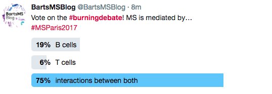
Both contestants had taken too many "peace-pills" rather than defend their corner, both threw in the towel to conclude that T and B cells were both (equally important) central to the problem of MS.

After a promising start, Prof.Hauser slipped on the canvas never recovered and started to verbally-punch himself with counter arguments such that Prof Hafler didn't have to bother defending himself or attacking the alternative view. Just as well, as he forgot his boxing gloves and came with the Immunological Karma Sutra of T and B cell interaction. 

With the thought of "Fake News" in both of the American contestants minds, could they defend any extreme position?


Perhaps there was fear of losing?

So much fun was had, that ProfG has gone for Round Two, and this time has persuaded ProfB (Lightweight) to take the challenge to Heinz Weindl (Heavy Weight). Today 


But to make it easier for Prof Weindl to accept the challenge, the loop-hole has been closed so that the T and B cells can be the problem as you have to explain how ocrelizumab works, which was the majority view expressed at ECTRIMS, when given the Choice.


The motion is
"To B or not-T-B that is the question"; the key pathogenic cell in MS is a B-cell and is independent of antigen presentation to T-cells"

Sounds like ProfB is in for a verbal kicking from the opponent and probably the audience. 

However, when the vote went live to twitter sphere after people had watched the videos and could not say both


Rather than a Boxing Match, maybe we should think of if it as a trial with the MS problem in the dock, 


There has been a game of Whodunnit called Cludedo


We have a victim the Ms. Oligodendrocyte has been murd**ed (killed). (If I use the word m******d, it triggers google to make this an adult site).

Where did the event take place?


We found the Body in the "Brain Room", but is that where the crime was plotted?

What was the killing weapon? 


Importantly Who is the killer and guilty, (Princess) B or Mr T? Are there any accomplices?


You will hear two theories/cases and the audience can be the Jury to "Acquit One" and "Find the other Guilty" as charged


Can we expect to see the "Chubbacca defence(deliberately confuse the jury rather than to factually refute the case of the other side) from the Weindl Team


The punishment will be the Death Penalty for the Guilty Party unless there can be rehabilitation.


The debate isn't being filmed, but we will rehearse the arguments from both side after the debate.

*****************************************************

Here are the slides (The Jokes have been removed)


As you can see the task given to ProfB was to argue that B cells can cause MS and this was not mediated by T cells and the activity due to B cells was not antigen presentation. 

This view was against what most people thought (about 60%). 

(a) The Key Idea presented was that the important subsets would come from looking at the response to therapy.

(b) It was argued by the audience that animal studies show that B cells present antigens to T cells in animals.

It was said because MOG T cell receptor transgenic (developed optic neuritis but not EAE) x MOG B cell receptor (immunoglobulin) transgenic mice (do not get EAE) get EAE means that B cells present antibody to T cells. 

This is not necessarily right as MOG T cell receptor transgenic mice, which get optic neuritis in both eyes, get sub-clinical spinal cord EAE. Add anti-MOG antibody and you get augmentation of disease and this can make mice develop clinical EAE, so no B cell presentation occurring.

Although B cells may well present antigens and there is evidence to support, this based on my extreme stance is irrelevant as animal models lack validity, because the CD20 depleting antibodies have a modest/marginal effect compared to that seen in humans.  We know animal models are T cell mediated. The aim here was not to discuss any of the animal work. MS is a human disease let's have a human based explanation

(c) Importantly the case was made that T cell depletion had a marginal effect. Now I could argue why the T cell depletion trials had not worked but I was attacking this view,

Again there was uproar by the T cell immunologists when it was suggested by that their much loved Th17 cells didn't do much in MS. 

This was contentious as expected. The audience and Prof W argued that IL-17A blockage didn't fail...ProfB's response was he didn't say it failed, he said it was no better than beta interferon, which is hardly a view that interleukin 17 was something magic. 

Activity of secukinumab, an anti-IL-17A antibody, on brain lesions in RRMS: results from a randomized, proof-of-concept study.Havrdová E, Belova A, Goloborodko A, Tisserant A, Wright A, Wallstroem E, Garren H, Maguire RP, Johns DR. J Neurol. 2016;263:1287-95.
The objective of this study was to assess the effect of secukinumab, a monoclonal antibody that inhibits interleukin (IL)-17A, on number of new active brain magnetic resonance imaging (MRI) lesions in subjects with relapsing-remitting multiple sclerosis (MS). Subjects (N = 73) were randomized 1:1 to secukinumab 10 mg/kg or placebo by intravenous infusion at weeks 0, 2, 4, 8, 12, 16, and 20. MRI scans were obtained within 30 days prior to randomization, on a monthly basis during the treatment period, and at study completion. The primary endpoint was the cumulative number of combined unique active lesions (CUAL) observed on brain MRI scans from week 4 to week 24. Compared with placebo, secukinumab non-significantly (MEANS THE TRIAL FAILED as the primary endpoint was not met) reduced the number of CUAL observed on 4-weekly MRI from week 4 to 24 (primary endpoint) by 49 % (95 % CI -10 to 77 %; P = 0.087) and significantly reduced the number of cumulative new gadolinium-enhancing T1 lesions by 67 % (31-84 %, P = 0.003). CUAL reductions were progressively greater from week 4 (1 %) to week 16 (49 %) and persisted until end-study (50 %). There were no serious adverse events; the adverse event rate was comparable to placebo (53 versus 49 %), although mild-to-moderate infection was somewhat more frequent (37 versus 23 %). This proof-of-concept study provides the first evidence that blocking IL-17A with an antibody may reduce MRI lesion activity in MS. Further studies are needed to confirm this finding and determine the magnitude of effect. 

The primary endpoint did not reach significance and so the study failed....That's how we do trials.

What does Beta interferon do?
Ann Neurol. 1994;36 Suppl:S113-4. This trial was monitored by both clinical and magnetic resonance imaging (MRI) methods. Clinical effect was a 30% reduction in relapse rate in the group treated with a high dose. The MRI activity rate was reduced by a median of 70% in both treatment groups.

De Stefano N, Curtin F, Stubinski B, Blevins G, Drulovic J, Issard D, Shotekov P, Gasperini C; IMPROVE Study Investigators.
Rapid benefits of a new formulation of subcutaneous interferon beta-1a in relapsing-remitting multiple sclerosis. Mult Scler. 2010; 16(7):888-92.

This study evaluated the efficacy of a new formulation of subcutaneous (sc) interferon (IFN)-β1a in relapsing—remitting multiple sclerosis (RRMS). Patients (n = 180) were randomized (2 : 1) to IFN-β1a or placebo for 16 weeks; all patients then received IFN-β1a for 24 weeks. Monthly brain MRI was performed. At week 16, the mean number of combined unique active (CUA) lesions was lower with IFN-β1a than with placebo (p < 0.001; 69% fewer lesions). The mean cumulative number of CUA lesions was already lower with IFN-β1a by week 4 (post hoc analysis; p = 0.015). The new formulation of sc IFN-β1a has rapid beneficial effects on MRI outcomes in RRMS

So anti-IL17 is hardly in the league of something like natalizumab.  

Were the T cell immunologists deluding themselves?

However, is it the inhibition of interleukin 17 actually due to Th17 effects? IL-17 is involved in B cell development and can enhance B cell proliferation and germinal center formation. Therefore it could effect memory B cell formation. I guess we will have to wait to see if this is found in psoriasis where the blocking antibody is being used. 

(d) The argument was made that Tcellers stop people understanding of how MS works because they view MS as a single cell type. This was clearly evident in the argument of ProfW

(d) An argument was that memory B cells and EBV was interlinked
The question was asked if you have antigen specific B cells, there must be antigen specific T cells. What are they doing? 

If the question was turned around and you ask a T celler, what are the antigen specific B cells doing? The answer may be who cares about B cells.

However, if EBV is driving the memory B cell activity, 
Does there need to be a T cell help as the virus can make naive  B cells proliferate and differentiate into memory B cells and this could be without T cells

There are testable hypothesis that come for these studies, 

I suspect the T cellers will continue with their T cell stuff as usual. This is fine meaning we can do it.

However, there was enough open-minded  people in the audience to see sense and they are the people who may be treating you.